1750 S Harbor Way Suite 100, Portland OR

About Us
Advanced Technology
State-of-the-Art Equipment for Superior Precision, Comfort & Results
At Malmquist Oral and Maxillofacial Surgery, we’re committed to providing the highest standard of care using the most advanced surgical and diagnostic technology available. Drs. Jay and Michael Malmquist continually invest in tools that enhance treatment accuracy, reduce discomfort, and improve long-term outcomes.
Explore the innovations that set our practice apart.
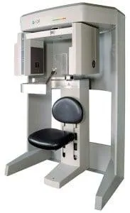
🧠 i-CAT 3D Imaging
Complete Diagnostic Clarity—In Seconds
Our i-CAT Cone Beam CT scanner delivers high-resolution, 3D images of your oral and facial structures in just seconds. This allows for unmatched precision in treatment planning and diagnosis.
Benefits of i-CAT imaging:
Optimizes implant placement and sizing
Identifies need for bone grafts or sinus lifts
Detects cysts, tumors, and jaw deformities
Accurately maps tooth position and vital structures
Reduces surgical risks and improves outcomes
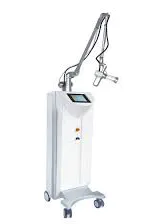
CO2 Laser
A Laser is a single wavelength beam of highly concentrated light energy. Medical surgery has been completely transformed by the laser. The laser has become a minimally invasive alternative to traditional surgical methods because it:
Is exact and predictable
Is highly efficient and produces little heat
Doesn’t require sterilization of the treatment area
Minimizes bleeding
Offers precise control over depth and extent of - incision
Requires no pain medication in some cases
Typically has little effect on the surrounding tissues
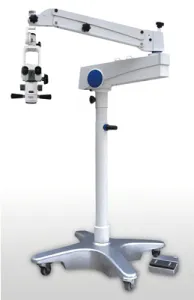
Surgical Microscope
Our implant treatment room is equipped with a special surgical microscope that enables Jay P Malmquist, DMD or Michael P Malmquist, DMD to provide minimally invasive procedures. This is accomplished with more precise surgical openings and fewer incisions, resulting in less trauma to the bone and soft tissue.
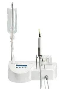
Piezosurgery
The three-dimensional ultrasound technology incorporated into this surgical device improves both the precision and predictability of various types of bone surgery.
The technology of controlled three-dimensional ultrasonic microvibrations opens up a new age in osseous surgery. The ultrasonic microvibrations of the Piezosurgery® device have been developed specifically for cutting bone tissue while minimizing trauma to the soft tissue.
This micrometric cutting action provides ultimate surgical precision and intra-operative sensitivity, while the selective cutting action maximizes safety for you and your patients. All of this is possible while operating with high intra-operative visibility and a blood-free surgical site.
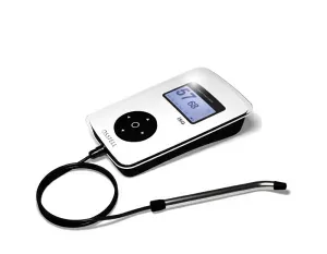
Osstell
It is critical that the bone be completely remodeled around the dental implants to maximize stability prior to attaching replacement teeth. The Osstell system will measure the stability of the implants, which enhances Jay P Malmquist, DMD or Michael P Malmquist, DMD‘s ability to determine more precisely the timing of the treatment sequence for each patient, and ultimately the predictability of the outcome.

5 Star Reviews
Our Clients Love Their Results
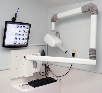
Digital Radiography
Digital Imaging
Dr. Malquist - Digital RadiographyJay P Malmquist, DMD and Michael P Malmquist, DMD choose carefully which and when radiographs are taken. There are many guidelines that we follow.
Radiographs allow us to see everything we cannot see with our own eyes. Radiographs enable us to detect cavities in between your teeth, determine bone level, and analyze the health of your bone. We can also examine the roots and nerves of teeth, diagnose lesions such as cysts or tumors, as well as assess damage when trauma occurs.
Dental radiographs are invaluable aids in diagnosing, treating, and maintaining dental health. Exposure time for dental radiographs is extremely minimal. Drs. Jay and Michael Malmquist utilizes Digital Imaging Technologies within the office. With digital imaging, exposure time is about 50 percent less when compared to traditional radiographs. Digital imaging can also help us retrieve valuable diagnostic information. We may be able to see cavities better. Digital imaging allows us to store patient images, and enables us to quickly and easily transfer them to specialists or insurance companies.
Digital X-Rays:
Digital X-rays offer more precision since we view the image on a computer monitor, instead of holding up a 35mm film up to the light. Digital X-rays results in 1/6th the radiation exposure to you.
In-House Discounts
We’re proud to offer a 5% discount on the day of surgery for payments made by cash or check.
This offer excludes dental implants and bone grafting procedures.
"Visit Our Clinic Today"
New Client Special - 20% OFF
Over 540+ Happy Customers
Frequently Asked Questions (FAQs)
Answers to Your Most Common Questions
What advanced technologies do you use in your practice?
At our practice, we use a comprehensive suite of advanced technologies to enhance diagnostic accuracy, surgical precision, patient comfort, and recovery times. These include i-CAT 3D imaging, CO₂ laser technology, a surgical microscope, Piezosurgery, the Osstell implant stability system, and digital radiography.
What is i-CAT 3D imaging, and why is it important?
i-CAT 3D imaging provides high-definition, three-dimensional scans of a patient's anatomy in real time. This allows Dr. Jay P. Malmquist and Dr. Michael P. Malmquist to evaluate bone structure, tooth orientation, and jaw joints with exceptional clarity. The imaging system helps identify bone deformities, cysts, tumors, or other pathology, and is especially useful in planning dental implants by determining exact positioning, proximity to vital structures, and whether procedures like bone grafting or sinus lifts are needed.
How does the CO₂ laser improve surgical procedures?
The CO₂ laser is a powerful tool that emits a focused beam of light energy, allowing for minimally invasive treatment of soft tissue conditions. It offers precision and predictability with minimal bleeding, little to no discomfort, and reduced need for anesthesia. It does not require the treatment area to be sterilized and has a minimal impact on surrounding tissues. We use the CO₂ laser for a variety of procedures such as crown lengthening, gum grafts, frenectomies, periodontal disease therapy, removal of excess gum tissue or skin imperfections, and laser-assisted dental implant surgery—all with virtually no bleeding or swelling.
What is a surgical microscope used for?
Our surgical microscope enhances visibility during procedures, allowing for greater accuracy and smaller incisions. This leads to less trauma to bone and soft tissue, quicker healing, and a more comfortable recovery. It plays a key role in achieving minimally invasive results during implant and soft tissue surgeries.
What is Piezosurgery, and how is it different from traditional techniques?
Piezosurgery uses three-dimensional ultrasonic microvibrations to cut bone with exceptional control while preserving nearby soft tissues. Unlike traditional surgical tools, this method allows for highly precise and safe osseous surgery. It provides clear visibility during the procedure, reduces bleeding, and ensures a more predictable outcome for both the surgeon and the patient.
How does digital radiography benefit me as a patient?
Digital radiography allows us to capture highly detailed images quickly while exposing patients to significantly less radiation compared to traditional X-rays. These images help us diagnose cavities, assess bone levels, examine tooth roots, and detect conditions such as cysts, tumors, or trauma-related damage. Because digital images can be stored electronically, they are easy to retrieve, share with specialists, and submit to insurance providers.
20% Off - First Visit
Schedule a Free Consultation
Experience Excellence in Oral and Maxillofacial Surgery
Address and E-Mail
Address
1750 S HARBOR WAY SUITE 100
PORTLAND, OR 97201
Get In Touch
Assistance Hours
Mon – Sat 9:00am – 8:00pm
Sunday – CLOSED

Copyright 2025. All Right Reserved.